developing a nano bio model system for rational design in nanomedicine

Developing a nano bio model system for rational design in nanomedicine
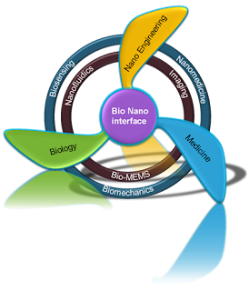

Nanomaterials with dimensions comparable with cell compartments, which are in the sub- micron or nano size domain, have unparalleled advantages in biomedical applications. The simple size compatibility allows us to use nanomaterials as small probes to decipher cellular machinery with minimal interference.. A wide range of nanomaterials including liposomes, polymeric micelles , quantum dots,carbon nanotubes , gold nanostructures (e.g. nanospheres, nanoshells, nanocages and nanorods, and nanowires  have shown exciting potential as imaging and sensing probes, drug and gene delivery carriers, and therapeutic agents. Although much progress has been made in this nano- bio direction, the translation of nanomedicine based on these nanostructures to a clinical setting has been hampered by the limited fundamental knowledge of the interactions between nanomaterials and biological systems. Understanding the interactions of nanomaterials with biological systems through systematic analysis is crucial to control and predict endocytosis as well as potential toxicity, and therefore enable rational design in nanomedicine. To establish this fundamental understanding, much effort has been spent using various organic and inorganic nanoparticles (NP). Organic NP, including liposomes, polymers and micelles have been widely used for nano- bio interaction studies. For instance, labeled liposomes were used to study the effect of ligand density on the binding effi ciency. Using liposomes that contain folate- conjugated polyethyleneglycolphosphatidylethanolamine, it was found that 0.5% folate produced the most effi cient binding of the liposomes to KB (a subline of the tumor cell line HeLa) carcinoma cells that overexpress the folate receptor. Using labeled spherical polymer particles;  the size effect on cellular uptake effi ciency of nanoparticles was reported. Labeled polymer nanoparticles have also been used to reveal the significant roles of particle shape in endocytosis and phagocytosis.. Discher and coworkers showed that fi lamentous polymer micelles persisted in the blood circulation up to 1 week after intravenous injection, about 10 times longer than the spherical counterparts. Using cationic poly(ethylene glycol)- based particles fabricated by top- down lithography, DeSimone and co- workers showed that rod- like particles have a higher cell internalization rate than the more symmetric counterparts. Using polystyrene particles as a model, Champion and  revealed an unexpected role of shape in phagocytosis. Recently, inorganic nanostructures, such as silica NP, gold NP, quantum dots (QD), carbon nanotubes (CNT) and nanowires (NW), have also gained more attention in studies of cellular interactions because of their unique material- and size- dependent physiochemical properties, which could be complementary to lipidor polymer- based nanoparticles. Fluorescently labeled silica NP with variable aspect ratio, size and surface functionalization were used to study NP translocation and cellular uptake and behavior (Huang et al. , 2010; Rancan et al. , 2012). Gold nanoparticles, including low- aspect-ratio gold nanorods and nanoshells, with strong luminescence and versatile surface chemistry have enabled researchers to investigate the effects of shape and surfactant coating on cellular uptake and cytotoxicity ;  Semiconductor QD with unique fl uorescence properties reveal a remarkable size and surface functionalization effect on cellular adsorption and endocytosis . CNT with intrinsic nearinfrared  and spontaneous Raman scattering have been tracked in live cells and live animals for studies of cellular interactions and CNT biodistribution in organisms. Very recently, NW were also used to explore cellular response. Magnetic nanowires were utilized to exploit their interaction with living cells in which cells were labeled with fluorescent probes . ZnO nanowires with intrinsic fluorescence have also been applied to the molecularly targeted imaging of cancer cells. Despite these advances, research along this direction faces several challenges
developing a nano bio model system for rational design in nanomedicine
First, current studies are challenged by lack of a strong intrinsic signal from the nanostructure. Although fluorescent agents are routinely used as labels for visualization of nanoscale drug carriers such as liposomes, polymer particles and copolymer micelles, they often suffer from the photo bleaching problem. More importantly, interpretation of fluorescence data can be further complicated because of dissociation of probes from the objects to be studied. Recent research by Cheng and co- workers revealed an unexpected release of lipophilic dyes from copolymer micelles  and poly(lactic- co-glycolic acid) nanoparticles  to lipid- rich structures during cellular uptake or blood circulation. To avoid labeling, near infrared (NIR) fluorescence  and spontaneous Raman scattering  have been used to study CNT in live cells and live animals. Intrinsic multiphoton luminescence  has been used to track gold nanorods and nanoshells in live cells and implanted tumors . Nevertheless, although the two- photon luminescence from gold nanostructures is as bright as the luminescence from quantum dots , more than 95% of the excitation energy is actually converted into heat, causing effective photo- toxicity to cells . Therefore, an intense and intrinsic optical signal with low damage potential is desired to follow a nanostructure in a biological system. Second, it is diffi cult to simultaneously control both the aspect ratio and surface chemistry of a nanostructure. For hydrogel particles fabricated by the soft lithography method, current studies have been restricted to non- targeting particles . For single- walled CNT, although various surface modifi cation schemes have been developed, it is hard to vary the diameter and/or control the length of the tube. Consequently, current studies have been focused on nanotubes with diameter limited to a few nanometers and length shorter than 200 nm. For gold nanorods, despite the versatile surface chemistry , it is difficult to prepare nanorods longer than 100 nm by the commonly used seeded growth method 

developing a nano bio model system for rational design in nanomedicine


Based on the above discussion, one would expect that an ideal model system for interrogating the cell- nanostructure interactions should meet three criteria: 1 an intensive and intrinsic signal that allows real- time visualization of single nanostructures with 3D submicron spatial resolution; 2 a surface capable of being modified in a mild condition to produce a controlled density of ligands or charges; 3 precise control of size as well as shape, that is aspect ratio for a rod shape, through fabrication or synthesis. Here, we introduce a newly developed excellent nano- bio model system based on functionalized silicon NW (SiNW) for study of the cellular response to 1D nanostructures. Our studies suggest that SiNW demonstrate three unique features meeting the criteria discussed above, including unparalleled dimension- control properties, intensive intrinsic NLO signals for imaging and flexible surface chemistry. The precise control of dimensions and aspect ratios of SiNW can be achieved through a metal nanocluster catalyzed chemical vapor deposition (CVD) method . In this chemical approach, monodisperse gold NP are used as the catalyst to control the diameter of SiNW.

developing a nano bio model system for rational design in nanomedicine


 Growth pressure and growth time are optimized to produce SiNW with desirable lengths ranging from a few hundred nanometers to tens of micrometers. We also discovered strong and stable third- order non- linear optical (NLO) signals (including four- wave mixing (FWM) and third- harmonic generation (THG) from SiNW, which, associated with deep penetration enabled by near- infrared or infrared pump beams and high 3D spatial resolution, can be used for both in vivo applications and label- free and noninvasive detection of the NW in biological experiments in real time. Additionally, SiNW with a native oxide layer on the surface can be easily functionalized based on well- studied silica surface chemistry. Collectively, we demonstrate that functionalized single crystalline SiNW can be employed to interrogate how a 1Dnanostructure interacts with cells in vivo and in vitro .

Comments

Popular posts from this blog

what is biosensor?

CT or computed tomography explained

how to prepare CuBTC?